Introduction
Ultrasound has long been a cornerstone of urologic imaging – it’s safe, real-time, and readily accessible. Now, artificial intelligence (AI) is gradually transforming urologic care by improving diagnostic precision and personalized treatment across many conditions[1]. While cutting-edge tools like MRI grab headlines, ultrasound remains the most widely used imaging modality in urology due to its convenience and lack of radiation[2]. The exciting news is that AI and ultrasound technologies are converging. As an experienced urologist, I’ve seen how even the best ultrasound can miss subtle details or depend on operator skill. AI offers a helping hand: it can analyze ultrasound images with superhuman consistency, highlight abnormalities, and even guide procedures. This means better care for patients – potentially earlier diagnoses, fewer unnecessary tests, and more targeted treatments delivered with confidence and compassion.
Patients understandably may feel anxious about medical tests and procedures. The integration of AI into ultrasound is ultimately about benefiting patients: making imaging more accurate, reducing invasive diagnostics, and personalizing therapy. In this educational overview, we’ll explore how AI is being applied in urologic ultrasound for both diagnostic purposes and interventional applications. We’ll discuss concrete examples – from AI finding hidden prostate tumors on an ultrasound, to guiding a novice’s hand during an ultrasound procedure. We’ll also highlight innovations from around the world (the United States and internationally) and touch on all forms of AI technologies being leveraged. Throughout, expect an evidence-based yet approachable explanation of this rapidly advancing field. By the end, you’ll understand how AI-powered ultrasound works, how it might affect your care as a patient, and what it means for healthcare professionals in urology.
AI Meets Ultrasound: A New Era of Imaging in Urology
Ultrasound imaging is invaluable in urology for visualizing the kidneys, bladder, prostate, and other organs. However, traditional ultrasound has well-known limitations: image quality can be variable with “speckle” noise; important structures may be hard to delineate; and interpretation is subjective and operator-dependent[3][4]. AI is poised to overcome many of these challenges. In simple terms, AI algorithms (including machine learning and deep learning) can be trained on large sets of ultrasound images to recognize patterns that correspond to specific diseases or anatomical landmarks. Unlike a human who might overlook a subtle shadow, a trained AI can sift through pixel data to find abnormalities that are hard to see. This translates to greater efficiency and fewer errors – one review noted that AI-supported ultrasound analysis can increase diagnostic efficiency and reduce subjective errors by automatically highlighting features for the clinician[4]. In practice, this means an AI might flag a tiny kidney stone or a faint lesion on the prostate ultrasound that a busy clinician could otherwise miss.
One of the biggest advantages of AI in ultrasound is its ability to standardize and automate tasks that currently vary between users. For example, measuring the size of an organ or finding the right angle for an image often depends on the operator’s expertise. AI tools can automate such measurements and provide real-time guidance. In fact, the European Association of Urology recently emphasized that new ultrasound advances, combined with training initiatives and AI, “promise to expand [ultrasound’s] role in precision diagnostics.”[5] By guiding less-experienced users to acquire quality images and by automatically analyzing those images, AI is effectively democratizing ultrasound imaging. Clinicians who are not imaging experts – say a primary care physician or a nurse in a rural clinic – can perform a scan with AI assistance and obtain diagnostic-quality results. This levels the playing field of care. “One of the biggest barriers to using ultrasound is having the right skills. But with AI-driven scan guidance…the user receives real-time, step-by-step feedback to maneuver the probe, almost like GPS, leading them to the optimal image,” explains Radhika Madhavan, an AI product manager at GE Healthcare[6]. In other words, AI is helping turn more healthcare providers into capable ultrasound users, which means patients can get timely imaging even outside major centers.
From a technical standpoint, AI in medical imaging can be split into two generations: earlier “machine learning” systems that relied on hand-crafted image features, and newer deep learning systems that automatically learn features from data[7]. The latter (often using convolutional neural networks) have been especially successful with ultrasound. They excel at tasks like recognizing organ boundaries, classifying tissue type, or detecting an anomaly – all in real-time as the ultrasound probe moves. For instance, if you’re getting a kidney ultrasound, an AI could simultaneously outline the kidney on the screen, measure its size, and alert the operator to any suspicious mass, all during the exam. The result is a more consistent and thorough exam, regardless of who is scanning. For patients, this means fewer “inconclusive” scans and a reduced likelihood of needing additional tests. Overall, this new era of AI-guided ultrasound promises faster, more accurate diagnoses while maintaining the safety and accessibility that make ultrasound so beloved in urology.
Diagnostic Applications of AI in Urologic Ultrasound
Prostate Cancer Detection and Prostate Health

One of the most impactful applications of AI in urologic ultrasound is in the detection of prostate cancer. Prostate cancer is common – about 1 in 8 men will be diagnosed in their lifetime – and early detection is key to effective treatment[8]. Traditionally, if a prostate cancer is suspected (for example, due to an elevated PSA blood test), the patient undergoes a biopsy guided by transrectal ultrasound (TRUS). During these biopsies, the urologist systematically takes 12-16 tissue samples from different regions of the prostate. This approach is somewhat blind; it’s been analogized to trying to find a flaw in a block of Swiss cheese by sampling through a few holes[9]. Not surprisingly, significant tumors can be missed – studies have shown standard systematic prostate biopsies can miss up to 50% of clinically significant cancers[10]. This is where AI can make a real difference for patients.
AI algorithms have been developed to analyze the ultrasound images of the prostate themselves to identify suspicious areas that might harbor cancer. A groundbreaking example is ProCUSNet, an AI tool developed at Stanford that examines ultrasound images taken during routine biopsy procedures[11]. ProCUSNet uses deep learning to localize areas of potential cancer on the standard B-mode ultrasound – essentially giving the urologist a “heatmap” of where cancer is likely. In a recent study, ProCUSNet was able to detect 82% of clinically significant prostate cancers and flagged 44% more lesions than expert radiologists could find when reviewing the same ultrasound images[12]. Impressively, the AI identified many high-grade tumors that the standard biopsy needles had initially missed; for patients who eventually had their prostate removed, nearly 30% had aggressive cancers that went undetected by conventional biopsy, but ProCUSNet caught signs of them[13]. What this means for patients is potentially fewer false assurances (“your biopsy was normal” when cancer is actually present) and fewer repeated procedures. An AI-enhanced ultrasound can help target the biopsy needles to the most suspicious areas in real time, improving the chances of finding any cancer that is there.
Another innovation in this sphere is the PCaVision system (by Angiogenesis Analytics), which is a computer-aided diagnostic platform using multiparametric ultrasound for prostate cancer. Unlike a standard ultrasound that only shows a gray-scale image, multiparametric ultrasound can include modes like elastography (which measures tissue stiffness) and contrast-enhanced ultrasound. The PCaVision system combines multiple ultrasound modes and then uses AI to generate real-time color-coded maps of the prostate highlighting regions suspicious for cancer[14]. It’s essentially an “AI second-opinion” during an ultrasound exam – as the urologist scans, the system is analyzing data under the hood and pointing out areas that deserve a closer look or a targeted biopsy. Clinical trials are underway to compare this AI-driven ultrasound approach to the current gold standard of MRI-targeted biopsies[15]. Early expectations are that such advanced ultrasound could perform nearly as well as MRI in finding significant cancers, which is exciting because ultrasound is cheaper, faster, and more accessible than MRI. For patients, an AI-enhanced ultrasound could mean faster diagnosis without always needing an MRI, especially in healthcare systems where MRI is hard to get. It also means that in the biopsy suite, the physician has extra guidance to ensure no important lesion is missed.
Beyond cancer detection, AI is helping with common prostate conditions like benign prostatic hyperplasia (BPH). BPH (an enlarged prostate) affects many men as they age and can cause urinary difficulties. Ultrasound is often used to measure the prostate’s volume, as a very enlarged prostate might warrant certain medications or procedures. Traditionally, measuring prostate volume on ultrasound involves the operator tracing the prostate’s outline or taking multiple dimensions – a process that can be time-consuming and prone to inter-user variation. Now, AI has stepped in to make this task almost instantaneous. In April 2025, an FDA-cleared tool called Clarius Prostate AI was introduced for a handheld ultrasound device, becoming the first AI tool specifically for prostate ultrasound[16][17]. This AI can automatically recognize the prostate on an ultrasound image, outline its borders, and calculate the prostate’s volume within seconds[18]. It even allows the clinician to input the patient’s PSA value, and then it automatically computes the PSA density (PSA level divided by prostate volume) on the spot[19]. PSA density is a useful indicator – a high PSA might be less concerning in a man with a very large prostate (because big prostates make more PSA in general), but more concerning in a man with a small prostate. By giving immediate PSA density results, the AI tool helps doctors identify which patients truly might be at higher risk for prostate cancer and which are likely just experiencing BPH[20]. For patients, this is a win because it enables earlier and more informed decision-making. A primary care doctor or urologist can perform a quick scan during an office visit, get the prostate size and PSA density, and decide right then if further tests or referrals are needed. This could spare patients from delays and extra appointments. And importantly, Clarius reports that over 95% of clinicians they surveyed believe such AI measurements save time and would allow them to see more patients in a day[21].
Kidney Ultrasound and Stone Detection

The kidneys and urinary tract are another area where AI in ultrasound is making waves. Ultrasound is often the first imaging test for kidney stones (urolithiasis), kidney masses, and hydronephrosis (swelling of the kidney due to urine blockage). However, ultrasound can sometimes miss small stones or have difficulty characterizing a mass. AI algorithms are tackling these issues head-on. In fact, researchers have achieved striking accuracy by applying AI to kidney ultrasound images. In one study, a team used a combination of traditional machine learning (SVM classifier) and neural networks to analyze kidney ultrasound scans – their system could automatically differentiate normal kidneys, kidney cysts, and kidney stones, with an accuracy of 98.1% in distinguishing cysts from stones on ultrasound[22]. In another project, investigators developed a deep learning model to detect renal calculi (stones) in ultrasound images despite the noisy background; their approach reached 98.8% accuracy in identifying kidney stones, outperforming conventional image processing methods[23]. These are remarkable numbers, suggesting that AI can tease out subtle features of stones (or absence thereof) that might elude the human eye. For patients suffering from suspected kidney stones, an AI-enhanced ultrasound could mean a more reliable diagnosis without having to resort to a CT scan in every case. (CT is very accurate for stones but involves radiation; if ultrasound can be made almost as accurate with AI, that’s a huge benefit in reducing radiation exposure and cost.)
AI is also proving useful in evaluating kidney masses. Ultrasound often finds lesions in the kidney (like a fluid-filled cyst versus a solid tumor), but sometimes it’s tricky to tell what a lesion is just by ultrasound brightness. Traditional practice might require a CT scan or MRI for clarification. AI pattern-recognition, however, can analyze the texture and features on ultrasound to help classify lesions. Early machine learning models have shown success in classifying kidney tumors on ultrasound versus benign findings[24]. This kind of AI assistance could non-invasively reassure a patient that a small lesion is likely a simple cyst (requiring no intervention) or flag that it has suspicious features requiring further workup.
In pediatric urology, one of the common issues is hydronephrosis – dilation of the kidney’s urine collecting system, often detected on prenatal ultrasound or in infants. Deciding on management (observation vs. surgery) partly depends on grading the severity of hydronephrosis on ultrasound. However, human grading is subjective and can vary between observers. AI is stepping in to bring objectivity. Researchers have applied deep learning models to automatically grade hydronephrosis severity from ultrasound images, which helps standardize assessments and potentially predict which children will need intervention. A systematic review noted that AI applications in renal ultrasound (including hydronephrosis evaluation) are a very promising area, though more validation is needed before they become routine[25]. As these models mature, parents of young patients may benefit from more consistent monitoring – for example, an AI could measure the exact size of the kidney’s pelvis on each scan and alert if there’s a significant worsening, prompting timely surgery and preventing kidney damage.
Importantly, the role of AI in kidney ultrasound is not just in detection, but also in reducing unnecessary procedures. If an AI can confidently identify a tiny kidney stone and gauge its size, a patient might avoid an invasive scope procedure just to locate the stone. Or if AI confirms a lesion is a benign cyst, the patient might avoid a biopsy. On the flip side, if something worrisome is found, AI can ensure it’s not overlooked, so the patient gets the needed treatment sooner. In essence, AI is acting as a tireless assistant, double-checking our ultrasounds for anything we should not miss when it comes to kidney health.
Bladder, Bladder Volume and Other Applications
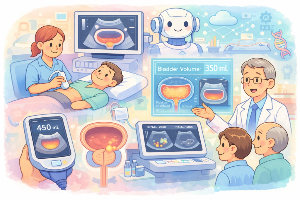
Ultrasound is frequently used to look at the bladder – often to measure how much urine it holds (bladder volume) or to check for post-void residual urine (how much urine is left after emptying). Accurately measuring bladder volume is important for patients with urinary retention, neurogenic bladder (e.g., due to spinal cord injury), or an enlarged prostate causing incomplete emptying. Conventionally, bladder volume is estimated by taking ultrasound measurements in two perpendicular planes and using a formula, or by using a dedicated bladder scanner device. Those devices can be costly, and manual measurements are a bit tedious. Here, AI shines by providing quick, automated readings. In January 2024, Clarius (the handheld ultrasound company) announced Bladder AI, an FDA-cleared tool that automatically measures bladder volume within seconds on their portable scanners[26][27]. The AI identifies the bladder in the ultrasound image, traces its outline, and calculates volume without the user having to do any mental math. According to the press release, Clarius Bladder AI was shown to “automatically and accurately measure bladder volume in seconds, saving time by improving workflow and efficiency.”[27] For a patient, this could mean a quicker, more comfortable assessment in the ER or clinic – the provider can immediately know if your bladder is holding too much urine, which might explain your discomfort or urinary issues.
The benefits go beyond just speed. Because the AI gives real-time feedback, it can guide the user to adjust the probe for the best view of the bladder. This ensures the volume calculation is based on a proper, centered image of the bladder. Such feedback is especially useful for nurses or clinicians who may not do ultrasounds frequently. Dr. Oron Frenkel, an emergency physician, noted that “Bladder AI removes the tedious steps of calculating bladder volume in my patients with urinary retention. It helps me quickly identify who needs a catheter….”[28]. This highlights a direct patient benefit: if the scan shows a large volume, the patient might need immediate catheterization to relieve retention; if the volume is low, maybe the issue is not retention and other causes of symptoms can be explored. By avoiding unnecessary catheterizations (which can be uncomfortable and carry infection risk), AI is helping improve patient comfort and safety. In fact, one study cited by the manufacturer found that using ultrasound (for bladder scanning) in clinical practice reduced the inappropriate use of urinary catheters, lowering patient risks and healthcare costs[29].
Beyond volume measurement, AI can also assist with detecting bladder abnormalities. For instance, although the definitive test for bladder cancer is a cystoscopy (a camera scope into the bladder), ultrasound sometimes is used to screen for large bladder tumors or thickening. AI image analysis could potentially flag subtle bladder wall irregularities or tumors on an ultrasound image, prompting earlier urology referral. Similarly, for testicular ultrasound (another urologic domain), AI is being explored to help differentiate between conditions like testicular torsion (an emergency) and epididymo-orchitis, or to characterize testicular masses. While these applications are still in development, they indicate how AI’s pattern recognition can extend to virtually any organ ultrasound.
Another noteworthy AI application is in fusion imaging. In prostate cancer, for example, we often fuse MRI images with ultrasound in real-time during biopsies to target suspicious areas seen on MRI. This typically requires external tracking devices and manual alignment. Researchers are now using AI to automatically align MRI and ultrasound images of the prostate in real time[30]. This AI-driven fusion can eliminate some of the human error in image co-registration and make advanced image-guided procedures more accessible without bulky equipment. Although this is behind-the-scenes technology, the end result for the patient is a smoother procedure – the doctor can see the MRI-defined tumor outline directly on the ultrasound screen with minimal fuss, ensuring the biopsy needle hits the mark.
In summary, AI is finding its way into many “odds and ends” of urologic ultrasound: from measuring how much urine your bladder holds, to helping distinguish a harmless cyst from a tumor, to fusing different imaging modalities for a comprehensive view. These developments all share a common goal: to make ultrasound exams more informative, reliable, and efficient, ultimately improving patient care.
AI-Enhanced Ultrasound-Guided Interventions
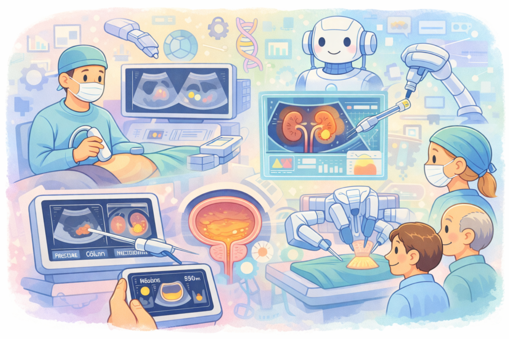
Beyond diagnostics, AI is also augmenting interventional ultrasound – that is, procedures where we use ultrasound imaging to perform a treatment or surgical task. In urology, common ultrasound-guided interventions include things like prostate biopsies (as discussed), kidney biopsies, placing drainage tubes in a kidney or abscess, and even guiding surgical procedures. AI can assist these interventions in several ways: by providing live guidance to the operator, automating certain steps, or improving accuracy and safety checks.
A prime example is again in prostate cancer care: AI targeting during prostate biopsies. Even when performing a standard biopsy, an AI like ProCUSNet can guide the urologist’s hand in real time: as the ultrasound shows the prostate, the AI might highlight a region with a red outline suggesting “biopsy here.” This effectively turns a systematic biopsy into a semi-targeted one, improving yield without changing the procedure workflow[31][32]. Looking ahead, as these AI tools become integrated into ultrasound machines, we can envision that every prostate biopsy will have an AI assistant in the background ensuring no suspicious area is overlooked. For the patient on the table, this could mean a higher likelihood that if cancer is present it will be found in one session, reducing the need for repeat biopsies (which are uncomfortable and carry some risk of bleeding or infection).
Another exciting area is robotic-assisted ultrasound interventions. There are research prototypes of AI-driven robotic systems that can perform or guide procedures using ultrasound imagery. For instance, a team from MIT developed an AI-guided device for central vein catheter placement in trauma settings[33][34]. In their system, the AI recognizes the target blood vessel on ultrasound and a robotic mechanism positions a needle, guiding a medic to successfully insert it even without prior training[35][36]. While this specific application is for emergency vascular access, the underlying concept can be applied in urology – imagine an AI-driven robot that could precisely insert a needle into a kidney stone under ultrasound guidance or place a tissue glue at a bleeding site. We’re not there yet for clinical urology, but these advances show the potential of AI + ultrasound + robotics to execute delicate tasks. Even without full automation, AI can assist human surgeons during ultrasound-guided procedures. For example, during a nephrostomy tube placement(inserting a tube into the kidney to relieve blockage), an AI could analyze the live ultrasound feed to identify the optimal entry point and angle, warn if the needle is veering toward adjacent organs, and confirm when the needle tip hits the fluid space. This would greatly help less experienced operators and could make bedside emergency procedures safer and more successful on the first attempt.
We already see simpler versions of this in clinical use. Certain ultrasound machines have AI features like cNerve that automatically detect and highlight nerves on ultrasound[37][38] – useful for anesthesiologists doing nerve blocks, but also relevant if a urologist is trying to avoid nerve injury or guide pain management injections. Another tool automatically labels anatomical structures (for instance, auto-labeling the kidney, liver, etc. on screen)[39], which can speed up procedures and reduce confusion. In prostate interventions, GE Healthcare’s new ultrasound platforms include AI-driven Prostate Volume Assist (PVA) that not only measures prostate size (as mentioned) but is integrated into guiding biopsy workflows[40]. It lets a urologist quickly get volume and then proceed to target sampling with fewer manual steps, streamlining the biopsy procedure.
Moreover, AI-based image fusion is an interventional aid. Consider MRI-ultrasound fusion biopsy: AI algorithms can automatically align the MRI-defined tumor with the real-time ultrasound, as discussed. This makes the intervention (targeted biopsy) more accurate without the operator spending time manually matching images or relying on cognitive fusion (essentially guessing the location on ultrasound by memory of MRI). Automatic fusion via AI could potentially shorten procedure time and improve targeting precision, leading to higher cancer detection rates per biopsy session. That’s a direct benefit to patients – a more efficient procedure with a higher diagnostic yield.
Another interventional domain is focal therapies for prostate or kidney tumors (like HIFU – high-intensity focused ultrasound – for prostate, or cryotherapy for small kidney tumors). These treatments often use ultrasound imaging to guide the therapy device. AI can assist by continuously tracking the target lesion on ultrasound, even if it moves slightly due to patient breathing or probe movement, and adjusting the therapy aim accordingly. It can also monitor changes (like tissue heating or ice ball formation) in the ultrasound image and alert the clinician if the therapy is not covering the entire tumor. Although these applications are still emerging, they illustrate how AI might function as a real-time co-pilot during therapy delivery.
In summary, AI in interventional ultrasound acts as an ever-vigilant guide and safety net. It augments the clinician’s skills: whether by highlighting where to put the needle, automating image alignment, or confirming procedural success criteria. The outcome for patients can be profound – interventions become safer, quicker, and more precise. For example, fewer needle passes to hit a target means less trauma and bleeding; precise localization of stones or tumors means more complete treatment in one session; and guided access in emergencies means potentially life-saving interventions delivered by generalists when specialists are not available. We are still in early days, but the trajectory is clear: AI will increasingly assist urologists in the OR and clinic to perform ultrasound-guided procedures with greater confidence and accuracy.
Benefits for Patients and Providers
The integration of AI into urologic ultrasound brings a host of benefits for both patients and healthcare professionals. At its heart, this technology is about improving outcomes and experience. Here are some of the key advantages:
- Earlier and More Accurate Diagnosis: By detecting subtle signs of disease on ultrasound that might be missed by the human eye, AI helps catch conditions at an earlier stage. For patients, this can mean finding a cancer while it’s still curable or identifying a kidney stone before it causes serious blockage. Enhanced accuracy also reduces the chance of misdiagnosis. For instance, if AI analysis of an ultrasound confidently labels a kidney mass as a simple cyst, the patient is spared the anxiety and risks of an unnecessary biopsy or surgery. In prostate cancer, tools like the AI we discussed can improve detection of significant tumors, potentially improving survival by ensuring timely treatment[12][13].
- Fewer Invasive Tests and Procedures: When ultrasound becomes more informative through AI, there’s less reliance on more invasive or costly diagnostics. A prime example is avoiding some diagnostic CT scans. Traditionally, a patient with suspected kidney stones might get a CT scan if the initial ultrasound was unclear. But if an AI-enhanced ultrasound clearly identifies a stone and pinpoints its size and location, a CT might be unnecessary. This spares the patient radiation exposure and lowers cost. Similarly, by better characterizing prostate nodules or kidney lesions on ultrasound, AI might reduce the number of biopsies done “just to be sure.” As noted in one analysis, AI-driven imaging can help distinguish benign from malignant findings, potentially “reducing unnecessary biopsies by distinguishing between BPH and cancer.”[41] For patients, every avoided invasive procedure is a relief in terms of comfort and safety.
- Personalized and Tailored Care: AI doesn’t just detect abnormalities; it can quantify them and integrate data for personalized decision-making. For example, automated prostate volume and PSA density calculation means the urologist can better stratify a patient’s cancer risk and tailor recommendations – maybe advising a biopsy or MRI for a high-density case, but just watchful waiting for a low-risk case[19][42]. In pediatric cases, consistent AI grading of hydronephrosis helps decide which children need surgery versus those who can be observed, avoiding overtreatment in some and undertreatment in others. Overall, AI provides quantitative, objective metrics that support more nuanced and individualized care plans.
- Time Savings and Efficiency: AI can take over repetitive tasks like measuring organ sizes, counting stones, or labeling anatomy, which frees up clinicians’ time during an exam. A task that might have taken 5-10 minutes (and full concentration) can be done in seconds by AI[21][18]. This not only allows the physician or sonographer to focus more on interacting with the patient (explaining findings, answering questions), but also means shorter scan times. Busy clinics benefit from this efficiency – more patients can be seen without long waits. From the patient’s perspective, a shorter ultrasound exam or procedure, and potentially getting results immediately, improves satisfaction. No one likes waiting nervously for measurements to be calculated or for a specialist to double-check images; AI provides instant second opinions and calculations.
- Enhanced Consistency and Confidence: By reducing operator dependence, AI ensures that no matter who performs the ultrasound, the key findings will be reliably identified. This consistency can especially benefit patients in areas with fewer specialists. A patient in a small town clinic can have confidence that their AI-assisted ultrasound is nearly as good as one reviewed by an expert in a university hospital – because the same trained AI model is working in the background. For providers, this takes away some pressure and uncertainty. Less experienced clinicians can proceed with more confidence that they aren’t missing something major, which in turn improves patient trust. Knowing that an AI has “double-checked” the scan and concurs (or flags something) gives both doctor and patient greater peace of mind.
- Improved Workflow and Reduced Burnout: On the provider side, AI can handle tasks like generating preliminary reports, annotating images, or even suggesting a diagnosis, which reduces administrative burden. For example, if after an AI-analyzed scan the system generates a draft report saying “Bladder volume: 300 mL, no significant residual, kidneys normal size, no stones detected,” the clinician only needs to verify and sign off. This reduces tedious documentation time. Studies have noted that lack of integration can be a barrier, but when AI is smoothly integrated, it can actually speed up workflow and let clinicians spend more time on direct patient care rather than staring at measurements and paperwork[41][43]. A faster workflow also means patients get results and next steps faster – less waiting for radiology reports on the back end.
- Better Patient Engagement and Understanding: AI can also help patients visualize and comprehend their condition. Some systems might display simplified graphics – for instance, coloring a prostate red in areas where suspicion is high – which a urologist can show to the patient to explain why a biopsy is needed. Or an automatic bladder volume readout can be shown to a patient to demonstrate “you’re retaining 400 mL of urine, which is why you feel discomfort.” This visual, data-driven approach can involve patients more in their care. Patients today are quite tech-savvy and often appreciate that advanced technology is being used for their benefit. Knowing that an AI is assisting (“a high-tech second opinion”) can be reassuring when explained properly. It is important, of course, for providers to communicate that AI is a tool and that human experts are still overseeing care, to maintain trust.
In summary, the fusion of AI with ultrasound in urology stands to make care more accurate, efficient, and patient-centered. As a urologist, I find that these tools, when used appropriately, augment our abilities – much like a stethoscope or MRI machine did in their times – ultimately leading to better patient outcomes. The key is that AI is not replacing the physician, but rather enhancing what we can do, allowing us to focus on the human aspects of care while the AI handles the number-crunching and pattern recognition in the background.
Education and Training Implications

The rise of AI in ultrasound doesn’t just help in direct patient care – it’s also changing how we train healthcare providers and how we educate patients. From a training perspective, AI-enabled ultrasound can serve as a virtual mentor or guide, particularly for those learning ultrasound skills. Traditionally, mastering ultrasound (for a urology resident, for example) requires many supervised scans to learn how to obtain the right views and interpret what’s on the screen. AI guidance systems accelerate this learning curve. They provide immediate feedback: “move the probe a bit to the left,” or “press a little harder to improve contact,” – akin to an expert looking over your shoulder. In fact, AI-guided scan systems have been likened to a “GPS for ultrasound imaging”[6], coaching the user to the correct probe position and angle. Studies on novice operators have shown that with AI assistance, even those with minimal training can acquire diagnostically useful images after a short training session, at times approaching the quality of experts. This is incredibly empowering – it means that a wider range of practitioners (from junior doctors to nurse practitioners) can be brought up to speed to perform basic ultrasound exams confidently. For urologists in training, an AI tool can highlight anatomy (like the ureter, renal pelvis, prostate boundaries) in real time, reinforcing their knowledge every time they scan a patient.
AI can also be used in simulation-based education. Imagine an ultrasound simulator that not only generates virtual patient scenarios but is equipped with an AI that reacts to the learner’s actions. If the learner’s probe placement is off, the AI simulator might indicate “image quality poor – try tilting the probe.” If a key finding is present on the image but the student doesn’t notice it, the AI could prompt, “do you see that shadowing in the kidney? What could that be?” This kind of interactive training makes learning ultrasound more engaging and thorough. It’s like having a personal tutor available 24/7. Over time, such training can produce clinicians who are adept at ultrasound much faster than with traditional methods alone, which again benefits patients (as there will be more skilled providers available).
For practicing clinicians, ongoing education is also supported by AI. Every time an AI highlights a finding, the clinician learns from it. For example, if an AI consistently marks small kidney stones that the clinician didn’t initially appreciate, the clinician’s own ability to spot stones improves. In essence, the AI is providing a continuous second opinion that one can learn from. In some AI software, after it makes a detection, it might also show which features were key (some research tools attempt “explainable AI” where it might, say, outline the texture patterns it found indicative of a tumor). This can deepen a clinician’s understanding of disease presentation on ultrasound.
From the patient education side, AI can help as well. When patients see visual aids – like color-coded risk areas on an ultrasound or an AI-generated 3D model of their prostate with marked spots – it can help them grasp the situation better than words alone. Some AI tools can even be integrated into patient-facing apps, potentially giving simplified explanations of ultrasound findings. For instance, a patient could receive an app message: “Your bladder scan today showed 50 mL of urine left after voiding, which is normal” – reassuring them and explaining in plain language. We are also seeing the emergence of AI chatbots trained on medical information that can answer patient questions post-visit, which could include explaining results of an ultrasound. While such uses are still experimental, they hint at a future where AI aids in closing the knowledge gap and anxiety gap for patients.
It’s worth noting that to fully realize these educational benefits, training the AI systems themselves on diverse operators and patients is important. AI should be seen as an adjunct in training, not a crutch – trainees should still learn the fundamentals of ultrasound physics and anatomy, using AI guidance as a support rather than completely relying on it. The goal is a well-rounded clinician who can operate with or without AI, but who uses AI to achieve expertise faster and maintain skills. From the patient perspective, education about AI’s role is needed too – some patients may worry “Is a computer doing my scan instead of a doctor?” It should be explained that the doctor/technologist is still performing the scan and making decisions; the AI is an assistant enhancing the process. When presented correctly, most patients find it reassuring that advanced AI technology is being used for their care, much like they appreciate a second specialist’s opinion.
In summary, AI in urologic ultrasound is not only transforming care delivery but also how we train providers and engage patients. It accelerates learning for clinicians, promotes consistency in practice, and can improve patient understanding of their health. This bodes well for the future – imagine more ultrasound-capable providers available in emergency rooms, clinics, and even remote areas, all thanks to AI-boosted training. Patients would have access to skilled ultrasound assessments no matter where they are, closing gaps in healthcare disparities (truly reflecting the “democratization” of ultrasound that AI can enable).
Global Innovations and Future Directions

The use of AI in ultrasound is a global phenomenon, with innovations arising from many corners of the world. Urologists and researchers internationally are collaborating to push this field forward, learning from each other’s successes. In the United States, we’ve seen startups and major companies driving clinical AI tools (e.g. the FDA-cleared prostate and bladder AI tools from Clarius Mobile Health, which is based in Canada but serves North America[44][45], and big imaging companies like GE Healthcare integrating AI across their ultrasound product lines). Europe is also at the forefront – for instance, the European Association of Urology’s 2025 congress highlighted ultrasound innovation including AI, such as the micro-ultrasound trials and the PCaVision AI system for prostate cancer mentioned earlier[46][14]. In the U.K., academic urologists have published narrative reviews and research on AI in urology (including ultrasound applications) to map current capabilities and challenges[47][48]. Asia, especially China, has contributed significantly on the research side; large datasets from hospitals there have been used to train deep learning models for kidney ultrasound diagnosis[49][50]. A 2023 systematic review from Chinese researchers analyzed dozens of AI-aided renal ultrasound studies, reporting high diagnostic accuracies but also noting the need for more robust validation[51][25]. Such cross-cultural research exchange means that an algorithm trained on data from one region can potentially benefit patients worldwide – an AI that learned from 10,000 ultrasound scans in Asia might accurately assist with scans in Europe or Africa, provided proper adjustments and validation.
One notable aspect of global innovation is how AI in ultrasound can bridge healthcare disparities. In many developing regions, advanced imaging like CT or MRI is scarce, but ultrasound machines (especially portable ones) are more available. AI can empower practitioners in those regions to utilize ultrasound to its fullest potential as a diagnostic tool, mitigating the lack of specialists. For example, consider a rural clinic in sub-Saharan Africa with a portable ultrasound device – if it’s equipped with an AI that can guide the user and identify, say, kidney obstruction or bladder stones, then patients there receive a higher level of care than would otherwise be possible on-site. There are global health initiatives looking to combine low-cost handheld ultrasounds with AI for precisely this reason: to provide high-quality imaging diagnostics in resource-limited settings. One prominent company in the handheld ultrasound space, Butterfly Network, has explored AI for automated interpretations (like lung ultrasound for pneumonia or bladder volume measures), aiming to deploy these in remote areas via telemedicine. Similarly, research groups in India have developed AI models for ultrasound detection of urinary stones and are looking to implement them in local hospitals where radiologist expertise might be limited[52][23]. It’s inspiring to see how international collaborations are forming – for instance, engineers in one country might develop the algorithm, clinicians in another validate it on their patients, and eventually regulators in multiple regions approve it for clinical use.
Regulatory bodies around the world are now catching up to these innovations. The U.S. FDA in the last couple of years has cleared multiple AI tools for ultrasound (the first being in cardiac ultrasound and now in urologic uses as well[16]). Europe’s CE marking process has likewise seen AI-enabled ultrasound devices gaining approval. These regulatory green lights are crucial, because they turn experimental innovations into real products that hospitals can buy and use. We can expect that as more data accumulates on safety and efficacy, regulators will become more comfortable with AI in clinical workflows, and approval times may shorten. It’s likely that within the next 5-10 years, AI features will be standard on most new ultrasound machines, just like color Doppler or 3D imaging became standard upgrades. In fact, if you walk into an ultrasound expo in 2025, many machines are being advertised for their AI capabilities – whether it’s auto-measuring organs, auto-finding lesions, or streamlining workflow.
The future directions in this field are exciting. One area of active development is improving AI’s explainability and robustness. Clinicians are more likely to trust and adopt AI if the system can explain why it’s flagging something (for example, highlighting the specific pixel pattern that suggests a tumor). Future AI might come with better user interfaces that show “AI confidence levels” or give a rationale (in simple terms) for its suggestions. This can also help with medicolegal acceptance – providing transparency that the doctor can review. Another direction is integration of multimodal data. Right now, most AI ultrasound tools focus just on the images. But imagine an AI that can take the ultrasound images plus clinical data (symptoms, lab results) and use all of it to generate an assessment. For instance, an AI might combine a renal ultrasound image with the patient’s blood creatinine, urinalysis, and medical history to predict the risk of that patient having a kidney obstruction or infection, not just identify structures. This holistic AI could then recommend “next steps” – perhaps suggesting “ultrasound findings plus data indicate high risk of kidney stone causing obstruction; consider urgent intervention.” This goes into the realm of clinical decision support, moving beyond just image interpretation.
In terms of research, we will see larger prospective trials of AI in urologic ultrasound. To truly prove benefit, we need studies that show using AI leads to better patient outcomes – e.g., randomized trials where one group gets AI-assisted ultrasound and the other standard care, and then measuring differences in diagnostic accuracy, time to diagnosis, complication rates, etc. Some trials like that are already starting (for example, evaluating AI vs MRI pathways for prostate cancer diagnosis[15]). These will give us valuable evidence on when and how to best deploy AI.
On the horizon, there are also futuristic ideas like using AI to control ultrasound probes autonomously (a robot could scan a patient’s kidney following a set protocol, guided by AI vision – useful in telemedicine or in space where an astronaut might need an ultrasound without a doctor present). Or creating “digital twins” of a patient’s organs – essentially 3D computer models built from ultrasound data, which AI continually updates and uses to run simulations (like predicting how a tumor will grow or how a stone will move). While such concepts sound like science fiction, they’re being explored in research labs and could become part of routine care in later decades.
Internationally, as these technologies mature, we expect to see convergence and collaboration. Urologists worldwide share the common goal of improving patient care. AI in ultrasound provides a new tool in that mission. Annual conferences now feature sessions on AI, with experts from different countries sharing experiences – what worked in one healthcare system, what challenges were faced in another. This helps create consensus guidelines on safely implementing AI. It’s likely that professional bodies (like the American Urological Association, European Association of Urology, etc.) will draft recommendations or best practices for using AI in ultrasound, ensuring quality control and addressing ethical considerations (like avoiding algorithmic bias against any population[43]).
In conclusion of the global perspective: innovation knows no borders, and the story of AI in urologic ultrasound is being written by a global community. Patients stand to benefit no matter where they live, as cutting-edge developments cross-pollinate internationally. The future looks bright – we anticipate smarter ultrasound machines that can do more and more on their own, integrated into clinical practice in a way that urologists and patients barely notice (except for the improved outcomes). The ultimate direction is that AI will make urologic care more precise, predictive, and personalized – a true leap forward akin to the introduction of ultrasound itself decades ago, but now supercharged for the 21st century.
Conclusion
AI in ultrasound is more than just a tech trend – it represents a compassionate advancement in how we diagnose and treat urologic conditions. As an urologist who has embraced these innovations, I can attest that the synergy of AI and ultrasound is already enriching patient care. We’ve journeyed through how AI helps spot prostate cancers hiding on routine scans, how it measures organs and detects stones with blinding speed and accuracy, and even how it guides needles and probes during critical interventions. Importantly, we’ve seen that these developments are grounded in real evidence and experience: from clinical trials to FDA clearances and beyond, AI in urologic ultrasound is not theoretical – it’s here and now, making a difference.
For patients, the message is reassuring. The incorporation of AI means your ultrasound exam is less likely to miss something important, and you might avoid extra tests or treatments thanks to more precise information upfront. It means if you live far from a big medical center, you can still have high-quality imaging done locally, with AI helping your providers. It means if you’re fearful about an upcoming procedure, there may be AI-driven tools in the room quietly working to make that procedure safer and faster. In the end, you remain in the care of trained medical professionals who use AI as a tool – much like a stethoscope or MRI – to do their jobs better. The empathy, judgment, and communication that a human doctor provides are irreplaceable; AI simply enhances the technical aspects, allowing the clinician to focus more on you.
For healthcare professionals, AI in ultrasound offers an opportunity to elevate our practice. It’s like having an extra pair of expert eyes and a tireless assistant who never gets fatigued. Of course, adopting these tools comes with a learning curve and the need to trust-but-verify the AI’s suggestions. There will be challenges – integrating new software, ensuring data privacy, and maintaining our own diagnostic skills. But as we have discussed, the rewards are great: improved accuracy, efficiency, and possibly even a restoration of some work-life balance by automating the grunt work. By embracing AI thoughtfully, we can enhance our precision without sacrificing the art of medicine. The tone among urologists and radiologists is increasingly optimistic, as early adopters share their success stories of AI catching a cancer earlier or cutting procedure time in half.
Looking ahead, the continued refinement of AI algorithms, accumulation of diverse training data, and rigorous clinical validation will further cement the role of AI in urologic ultrasound. We’ll likely see guidelines emerging on how best to use (and not use) these tools, and curricula adapting to teach new physicians about AI fluency. Importantly, patients will play a role in this evolution too – their comfort and trust in AI-assisted care will grow as the technology proves itself reliable and beneficial in real-world settings. Ongoing dialogue with patients about these advances will remain crucial, to ensure they feel informed and empowered in their care.
In conclusion, the use of AI in ultrasound – particularly in urology – exemplifies the fusion of cutting-edge innovation with compassionate care. It leverages the latest in computer science to serve one of the oldest goals in medicine: seeing clearly inside the body to diagnose and heal. The diagnostic and interventional applications we’ve explored show how far we’ve come, and the global momentum suggests we’re only at the beginning of this transformation. As we proceed, the guiding principle will be keeping the patient’s benefit at the center. With that compass, AI will undoubtedly be a force for good – a true game-changer in ultrasound imaging that helps save lives, reduce suffering, and improve the quality of care for countless patients in the years to come.
References:
- Samenezhad S, Rafighi D. The role of artificial intelligence in advancing urologic care: From diagnostics to therapeutics. Surgery in Practice and Science. 2026;24:100322[1].
- Pérez Ardavín J. Ultrasound innovation in urology: What’s new in 2025? European Association of Urology (EAU) News. Oct 1, 2025[2][5].
- Liang X, et al. Artificial intelligence-aided ultrasound in renal diseases: a systematic review. Quant Imaging Med Surg. 2023;13(6):3311-3327[4][25].
- Stanford Medicine Department of Radiology. AI Tool Boosts Detection of Clinically Significant Prostate Cancer with Routine Ultrasound (News Release). Jan 2025[12][13].
- Clarius Mobile Health. FDA Clears New Clarius Prostate AI to Help Physicians Assess and Diagnose Common Urologic Conditions in Minutes (Press Release). Apr 25, 2025[17][18].
- Clarius Mobile Health. Clarius Adds AI-Driven Automatic Bladder Volume Measurements to Wireless Handheld Ultrasound Scanners (Press Release). Jan 9, 2024[27][28].
- Rahman ME, et al. Artificial Intelligence in Urolithiasis Imaging and Intervention: A Narrative Review. Cureus. 2025;17(11):e97716[22][23].
- GE HealthCare. AI-Guided Ultrasound: From Innovation to Impact (Insights Article). Jan 2, 2025[53][6].
- Byram Healthcare. The Rise of Artificial Intelligence in Urology (Blog). 2024[41][43].
[1] The role of artificial intelligence in advancing urologic care: From diagnostics to therapeutics – ScienceDirect
[2] [5] [14] [15] [30] [46] Ultrasound innovation in urology: What’s new in 2025? – Uroweb
[3] [4] [7] [25] [49] [50] [51] Artificial intelligence-aided ultrasound in renal diseases: a systematic review – Liang – Quantitative Imaging in Medicine and Surgery
[6] [53] AI-Guided Ultrasound: From Innovation to Impact | GE HealthCare (United States)
[8] [9] [10] [11] [12] [13] [31] [32] AI Tool Boosts Detection of Clinically Significant Prostate Cancer with Routine Ultrasound | Radiology | Stanford Medicine
[16] [17] [18] [19] [20] [21] [42] [44] FDA Clears New Clarius Prostate AI To Helps Physicians Assess and Diagnose Common Urologic Conditions in Minutes | Clarius | Press Release
[22] [23] [24] [47] [48] [52] Artificial Intelligence in Urolithiasis Imaging and Intervention: A Narrative Review of Current Applications, Barriers, and Future Directions – PMC
[26] [27] [28] [29] [45] Clarius Adds AI-Driven Automatic Bladder Volume Measurements to its Wireless Handheld Ultrasound Scanners Designed for Acute Care, Urology, and Nursing
[33] [34] [35] [36] Artificial Intelligence–Guided Ultrasound Intervention Device | MIT Lincoln Laboratory
[37] [38] [39] [40] Verisound AI | GE HealthCare (United States)
[41] [43] The Rise oArtificial Intelligence in Urologic Ultrasound: Enhancing Diagnosis and Interventionf Artificial Intelligence (AI) in Urology